Discover how advanced microfabrication technologies are revolutionizing organ-on-chip development, enabling more predictive drug discovery and personalized medicine research.
Understanding the Foundation of Organ-on-Chip Technology
Organ-on-chip technology represents a transformative approach to modeling human physiology in vitro, bridging the gap between traditional cell culture and animal models. These microfluidic devices recreate the mechanical, chemical, and biological microenvironment of living organs, enabling researchers to observe cellular behavior under physiologically relevant conditions. By incorporating dynamic fluid flow, mechanical forces, and tissue-tissue interfaces, organ-on-chip systems provide unprecedented insights into human disease mechanisms and drug responses.
The foundation of successful organ-on-chip development lies in understanding the complex interplay between material properties, cell biology, and engineering design. Researchers must consider factors such as oxygen tension, shear stress, nutrient gradients, and cell-cell communication when designing these sophisticated platforms. Advanced microfabrication techniques enable precise control over these parameters, creating reproducible environments that mirror in vivo conditions. This level of precision is particularly valuable for pharmaceutical R&D labs seeking to reduce development timelines and improve the predictive accuracy of preclinical testing.
As drug discovery pipelines increasingly demand more human-relevant models, organ-on-chip technology offers a pathway toward personalized medicine and more efficient therapeutic development. These platforms can incorporate patient-derived cells, enabling researchers to study individual disease phenotypes and drug responses. The integration of automation and standardization in organ-on-chip fabrication is helping to transition this technology from specialized research tools to scalable platforms suitable for high-throughput screening applications.
Precision Microfabrication Techniques That Define Quality
The quality of organ-on-chip devices depends heavily on the precision and reproducibility of microfabrication techniques employed during manufacturing. Photolithography remains the gold standard for creating microfluidic channel patterns, offering submicron resolution and excellent repeatability across production batches. Soft lithography using polydimethylsiloxane (PDMS) has become particularly popular due to its optical transparency, gas permeability, and biocompatibility. However, emerging techniques such as two-photon polymerization and laser-based fabrication are pushing the boundaries of what's achievable in terms of geometric complexity and feature resolution.
Advanced fabrication platforms like the Nanoscribe Quantum X enable researchers to create intricate three-dimensional microarchitectures with unprecedented precision. These high-resolution systems utilize 3D laser lithography to produce complex scaffolds and microfluidic geometries that would be impossible to achieve with traditional planar fabrication methods. The ability to incorporate multiple length scales—from nanometer-scale surface features to millimeter-scale device dimensions—is essential for recapitulating the hierarchical organization found in native tissues.
Quality control in microfabrication extends beyond dimensional accuracy to include surface properties, optical clarity, and functional performance. Researchers must validate that channel dimensions remain consistent across devices, that surfaces support appropriate cell adhesion, and that fluid dynamics match design specifications. Automated inspection systems and standardized testing protocols help ensure that each device meets the stringent requirements necessary for reliable biological experiments. This attention to fabrication quality is what separates research-grade prototypes from platforms capable of supporting rigorous pharmaceutical development programs.
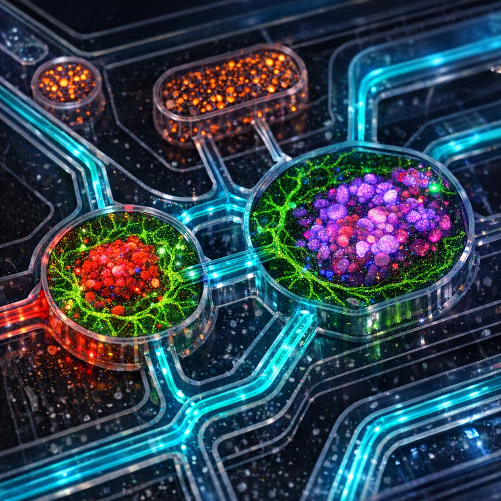
Integrating Biocompatible Materials and Cell Systems
The selection of biocompatible materials is a critical decision point in organ-on-chip fabrication, directly impacting cell viability, function, and experimental outcomes. While PDMS has dominated the field due to its favorable properties, researchers are increasingly exploring alternatives such as thermoplastics, hydrogels, and composite materials that offer enhanced control over mechanical properties and reduced small-molecule absorption. Materials must not only support cell survival but also provide appropriate mechanical stiffness, surface chemistry, and permeability to match the target tissue environment.
Integrating living cells into microfluidic devices requires careful consideration of cell seeding methods, culture conditions, and long-term maintenance strategies. Single-cell dispensing technologies, such as those offered by platforms like UP.SIGHT and CELLMATE, enable precise placement of specific cell types within defined chip regions. This level of control is essential for creating co-culture systems that replicate tissue-tissue interfaces, such as the vascular-parenchymal barriers found in organs. Automated cell handling reduces variability and improves throughput, making it feasible to establish complex multi-organ systems with reproducible cellular compositions.
The choice of cell source—whether immortalized cell lines, primary cells, or stem cell-derived cells—significantly influences the physiological relevance of organ-on-chip models. Patient-derived induced pluripotent stem cells (iPSCs) offer the exciting possibility of personalized disease modeling, though they require sophisticated differentiation protocols and quality control measures. Regardless of cell source, maintaining appropriate phenotypes under microfluidic culture conditions often requires optimization of media formulations, flow rates, and biomechanical stimulation. Successfully integrating biocompatible materials with robust cell systems transforms organ-on-chip devices from engineering accomplishments into powerful research tools that can genuinely improve drug discovery workflows.
Microfluidics Design Principles for Physiological Relevance
Effective microfluidics design for organ-on-chip applications requires translating physiological principles into engineered fluid dynamics. Researchers must consider parameters such as shear stress, fluid velocity, residence time, and mass transport to recreate the dynamic environment cells experience in vivo. For vascular models, this means implementing flow rates and channel geometries that generate physiologically relevant shear stress levels, typically in the range of 0.5-20 dynes/cm². For epithelial tissues, designing appropriate air-liquid interfaces or chemical gradients may be more critical than fluid flow itself.
The architecture of microfluidic channels determines not only fluid dynamics but also nutrient delivery, waste removal, and the establishment of biochemical gradients. Multi-layer designs can separate vascular compartments from tissue compartments, mimicking the organization of living organs while enabling independent control of each microenvironment. Porous membranes incorporated between layers facilitate cell-cell communication and molecular transport while maintaining distinct fluidic paths. These design elements are essential for studying processes such as drug absorption, immune cell migration, and metastatic invasion that depend on interactions across tissue boundaries.
Advanced microfluidics design increasingly incorporates sensors and real-time monitoring capabilities to track tissue function and drug responses dynamically. Integrated electrodes can measure barrier integrity in real-time, while optical access enables high-resolution imaging of cellular structures and molecular reporters. The challenge lies in balancing design complexity with practical usability—overly complicated systems may offer more physiological features but can be difficult to operate reliably and scale for higher-throughput applications. The most successful organ-on-chip platforms strike a balance between physiological relevance and experimental accessibility, making them valuable tools for both academic researchers and pharmaceutical R&D teams seeking to improve the predictive power of their preclinical models.
Validation Strategies and Quality Control Considerations
Rigorous validation is essential for establishing confidence in organ-on-chip platforms as predictive tools for drug discovery and toxicology assessment. Validation strategies typically proceed through multiple stages, beginning with characterization of device fabrication consistency, followed by assessment of cellular function, and culminating in comparison with in vivo responses or clinical outcomes. Researchers must demonstrate that their organ-on-chip models recapitulate key tissue-specific functions, respond appropriately to positive and negative controls, and generate reproducible results across independent experiments and operators.
Quality control considerations span the entire organ-on-chip workflow, from raw material sourcing through cell culture protocols to data analysis methods. Establishing standard operating procedures (SOPs) for device preparation, cell seeding, culture maintenance, and endpoint measurements is critical for achieving reproducibility. Implementing automated systems for cell dispensing, media exchange, and environmental control reduces operator-dependent variability. Platforms that integrate workflow management software help research teams maintain consistency across complex multi-step protocols, ensuring that data quality meets the standards required for regulatory submissions or publication in high-impact journals.
As organ-on-chip technology matures, the field is moving toward standardization of validation criteria and performance benchmarks. Industry consortia and regulatory agencies are beginning to establish guidelines for what constitutes a sufficiently validated model for specific applications. For pharmaceutical companies, demonstrating that an organ-on-chip platform can correctly classify known toxicants or predict clinical drug responses is essential for justifying its incorporation into development pipelines. Academic researchers benefit from these validation frameworks as well, as they provide clear targets for model development and facilitate comparison across different platforms. By maintaining high standards for validation and quality control, the organ-on-chip community is building the foundation for these technologies to transform how we approach drug discovery and personalized medicine.